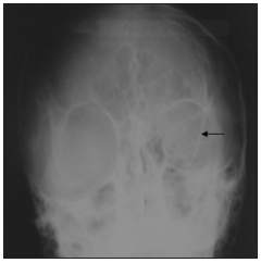
Neurofibromatosis with pulsating exophthalmos
Main Article Content
Abstract
Background: Neurofibromatosis (NF) is a neurocutaneous disorder which involves many organs in the body. There are two types: NF-1 and NF-2. Orbital manifestation is a rarity in NF-1, and it involves dysplasia of the sphenoid bone resulting in herniation of the temporal lobe and subarachnoid space into the orbit culminating in pulsating exophthalmos.
Aim: To highlight the clinical presentation and radiological investigation of this rare ocular manifestation of NF-1.
Methods: A case report.
Results: The case of a 20-year-old male student presenting with a pulsating right eye swelling of about 17- year duration is presented. There was a family history of a first-degree relative with multiple skin swellings. Plain skull radiograph and cranial computed tomography (CT) scan were done and both revealed absence of the right sphenoid bone with herniation of the right temporal lobe and cerebrospinal fluid space into the right orbit. The patient was subsequently lost to follow-up.
Conclusion: Pulsating exophthalmos is a complication of sphenoid dysplasia, a rare component of NF-1. Plain skull radiograph and cranial CT scan are two important radiological imaging modalities for investigating patients with such presentation.
Downloads
Article Details
The journal grants the right to make small numbers of printed copies for their personal non-commercial use under Creative Commons Attribution-Noncommercial-Share Alike 3.0 Unported License.
References
1. Fortman BJ, Kuszyk BS, Urban BA, Fishman EK. Neurofibromatosis type 1: A diagnostic mimicker at CT. Radiographics 2001;21:601‑12.
2. Sutton D, Stevens J, Miszkiel K. Intracranial lesions (1). In: Sutton D, editor. Textbook of Radiology and Imaging. 7th ed. Philadelphia: Elsevier, 2003; 1723‑66.
3. Gunny RS, Kling Chong WK, Paediatric neuroradiology. In: Adam A, Grainger RG, Dixon AK, Allison DJ, editors. Grainger and Allison Diagnostic Radiology: A Textbook of Medical Imaging. 5th ed. Philadelphia: Elsevier, 2008; 1653‑701.
4. Baskurt E. Cranial and spinal imaging. In: Gay SB, Woodcock RJ, editors. Radiology Recall. 2nd ed. Philadelphia: Lippincott Williams and Wilkins, 2008; 700‑70.
5. Dahnert W. Radiology Review Manual. 6th ed. Philadelphia: Lippincott Williams and Wilkins, 2007; 316‑9.
6. Levy AD, Patel N, Dow N, Abbott RM, Miettinen M, Sobin LH. From the archives of the AFIP: Abdominal neoplasms in patients with neurofibromatosis type 1: Radiologic‑pathologic correlation.
Radiographics 2005;25:455‑80.
7. Patronas NJ, Courcoutsakis N, Bromley CM, Katzman GL,
MacCollin M, Parry DM. Intramedullary and spinal canal tumors in
patients with neurofibromatosis 2: MR imaging findings and correlation
with genotype. Radiology 2001;218:434‑42.
8. Bognanno JR, Edwards MK, Lee TA, Dunn DW, Roos KL, Klatte EC.
Cranial MR imaging in neurofibromatosis. AJR Am J Roentgenol
1988;151:381‑8.
9. Bunin GR, Needle M, Riccardi VM. Paternal age and sporadic
neurofibromatosis 1: A case‑control study and consideration of the
methodologic issues. Genet Epidemiol 1997;14:507‑16.
10. Jacquemin C, Bosley TM, Liu D, Svedberg H, BuhaliqaA. Reassessment
of sphenoid dysplasia associated with neurofibromatosis type 1. AJNR
Am J Neuroradiol 2002;23:644‑8.
11. Jacquemin C, Bosley TM, Svedberg H. Orbit deformities in craniofacial
neurofibromatosis type 1. AJNR Am J Neuroradiol 2003;24:1678‑82.
12. Chung EM, Specht CS, Schroeder JW. From the archives of the
AFIP: Pediatric orbit tumors and tumorlike lesions: Neuroepithelial
lesions of the ocular globe and optic nerve. Radiographics
2007;27:1159‑86.
13. Rogers LF, Auringer ST. The congential malformation syndromes:
Osteochondrodysplasias, dysostoses and chromosomal disorders. In:
Juhl JH, Crummy AB, Kuhlman JE, Paul LW, editors. Paul and Juhl’s
Essential of Radiologic Imaging. 7th ed. Philadelphia: J B Lippincott
company, 1998.
14. Akadiri O, Jackson I. Craniofacial neurofibromatosis type 1: Clinical
features, challenges of management, and a report of 2 Nigerian
patients. Int J Head Neck Surg 2008;3:1-7.
15. Wakely SL. The posterior vertebral scalloping sign. Radiology
2006;239:607‑9.
16. Chapman S, Nakielny R. Aids to Radiological Differential Diagnosis.
4th ed. Edinburg: Elsevier, 2003; 375‑96.
17. Hope RA, Longmore JM, McManus SK, Wood‑Allum CA. Oxford
Handbook of Clinical Medicine. 4th ed. Oxford: Oxford University
Press, 1998; 474‑5.
18. Cai W, Kassarjian A, Bredella MA, Harris GJ, Yoshida H, Mautner VF,
et al. Tumor burden in patients with neurofibromatosis types 1 and
2 and schwannomatosis: Determination on whole‑body MR images.
Radiology 2009;250:665‑73.


