Measurement of thoracic and lumbar pedicle dimensions in Nigerians using computed tomography
Main Article Content
Abstract
Background: Pedicle screws are often used to stabilise the spine. They afford the benefit of a three-column control of the spine. The technique of pedicle screw insertion is familiar and has a well-documented safety profile during lumbar and thoracic spinal surgery. However, complications such as cerebrospinal fluid leakage due to pedicle screw misplacement, neurological irritation and pedicle penetration may occur. Therefore, knowledge of the dimensions of spinal pedicles is necessary for the fixation of pedicular screws to avoid possible complications.
Aims: The aim of this study was to determine the maximal diameter and axial length of thoracic and lumbar pedicles in a homogenous African population using computed tomography (CT). This would establish normative data on the average size of pedicle screws that would be required during the surgery, hence maximising pull-out strength while reducing the possibility of revision of the pedicle screw placement.
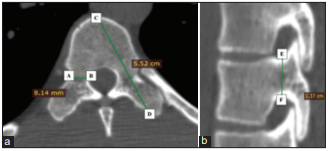
Methods: It is a retrospective study where the transverse pedicle width, axial pedicle length and sagittal pedicle width of T1–L5 were measured on 100 patients; 50 males, 50 females with normal spinal architecture using a 128-slice Toshiba CT scanner.
Results: The mean axial length in the thoracic and lumbar vertebrae ranged from 31.76 ± 2.92 mm (T1) to 43.02 ± 3.32 mm (T12) and from 45.07 ± 2.40 mm (L5) to 46.32 ± 2.28 mm (L3), respectively. The mean TPW at the thoracic and lumbar vertebrae ranged from 4.53 ± 0.69 (T4) to 7.78 ± 1.31 mm (T12) and from 6.81 ± 1.25 mm (L1) to 12.95 ± 1.49 mm (L5), respectively. The mean sagittal diameter of thoracic and lumbar vertebrae ranged from 5.78 ± 1.07 mm (T1) to 10.98 ± 1.37 (T12) and from 9.51 ± 1.31 mm (L2) to 9.78 ± 1.61 (L4), respectively.
Conclusion: The dimensions of thoracic and lumbar pedicles measured in this study vary with those obtained from other populations. This strengthens the case for customising the existing range of spinal pedicle screws according to local population characteristics.
Downloads
Article Details
The journal grants the right to make small numbers of printed copies for their personal non-commercial use under Creative Commons Attribution-Noncommercial-Share Alike 3.0 Unported License.
References
1. Lee KD, Lyo IU, Kang BS, Sim HB, Kwon SC, Park ES. Accuracy of pedicle screw insertion using fluoroscopy‑based navigation‑assisted surgery: Computed tomography postoperative assessment in 96 consecutive patients. J Korean Neurosurg Soc 2014;56:16‑20.
2. Urrutia VE, Eliozondo OR, De La Garza CO, Guzmán LS. Morphometry of pedicle and vertebral body in a Mexican population by CT and fluroscopy. Int J Morphol 2009;27:1299‑303.
3. Chapman JR, Harrington RM, Lee KM, Anderson PA, Tencer AF, Kowalski D. Factors affecting the pullout strength of cancellous bone screws. J Biomech Eng 1996;118:391‑8.
4. Ruf M, Harms J. Pedicle screws in 1‑ and 2‑year‑old children: Technique, complications, and effect on further growth. Spine (Phila Pa 1976) 2002;27:E460‑6.
5. Krag MH, Beynnon BD, Pope MH, Frymoyer JW, Haugh LD, Weaver DL. An internal fixator for posterior application to short segments of the thoracic, lumbar, or lumbosacral spine. Design and testing. Clin Orthop Relat Res 1986;203:75-98.
6. Roy‑Camille R, Saillant G, Mazel C. Internal fixation of the lumbar spine with pedicle screw plating. Clin Orthop Relat Res 1986;203:7‑17.
7. Youkilis AS, Quint DJ, McGillicuddy JE, Papadopoulos SM. Stereotactic navigation for placement of pedicle screws in the thoracic spine. Neurosurgery 2001;48:771‑8.
8. Vaccaro AR, Rizzolo SJ, Balderston RA, Allardyce TJ, Garfin SR, Dolinskas C, et al. Placement of pedicle screws in the thoracic spine. Part II: An anatomical and radiographic assessment. J Bone Joint Surg Am 1995;77:1200‑6.
9. Cheung KM, Ruan D, Chan FL, Fang D. Computed tomographic osteometry of Asian lumbar pedicles. Spine (Phila Pa 1976) 1994;19:1495‑8.
10. Hou S, Hu R, Shi Y. Pedicle morphology of the lower thoracic and lumbar spine in a Chinese population. Spine (Phila Pa 1976) 1993;18:1850‑5.
11. Zindrick MR, Wiltse LL, Doornik A, Widell EH, Knight GW, Patwardhan AG, et al. Analysis of the morphometric characteristics of the thoracic and lumbar pedicles. Spine (Phila Pa 1976) 1987;12:160‑6.
12. Suk SI, Kim WJ, Lee SM, Kim JH, Chung ER. Thoracic pedicle screw fixation in spinal deformities: Are they really safe? Spine (Phila Pa 1976)
2001;26:2049‑57.
13. Guyer DW, Yuan HA, Werner FW, Frederickson BE, Murphy D. Biomechanical comparison of seven internal fixation devices for the
lumbosacral junction. Spine (Phila Pa 1976) 1987;12:569‑73.
14. Masferrer R, Gomez CH, Karahalios DG, Sonntag VK. Efficacy of pedicle screw fixation in the treatment of spinal instability and failed back surgery: A 5‑year review. J Neurosurg 1998;89:371‑7.
15. Steffee AD, Biscup RS, Sitkowski DJ. Segmental spine plates with pedicle screw fixation. A new internal fixation device for disorders of the lumbar and thoracolumbar spine. Clin Orthop Relat Res 1986;203:45-53.
16. Castro WH, Halm H, Jerosch J, Malms J, Steinbeck J, Blasius S. Accuracy of pedicle screw placement in lumbar vertebrae. Spine (Phila Pa 1976) 1996;21:1320‑4.
17. Weinstein JN, Rydevik BL, Rauschning W. Anatomic and technical considerations of pedicle screw fixation. Clin Orthop Relat Res 1992;284:34-46.
18. Maaly MA, Saad A, Houlel ME. Morphological measurements of lumbar pedicles in Egyptian population using computerized tomography and cadaver direct caliber measurements. Egypt J Radiol Nucl Med 2010;41:475‑81.
19. Austin MS, Vaccaro AR, Brislin B, Nachwalter R, Hilibrand AS, Albert TJ. Image‑guided spine surgery: A cadaver study comparing conventional open laminoforaminotomy and two image‑guided techniques for pedicle screw placement in posterolateral fusion and nonfusion models. Spine (Phila Pa 1976) 2002;27:2503‑8.
20. Laine T, Lund T, Ylikoski M, Lohikoski J, Schlenzka D. Accuracy of pedicle screw insertion with and without computer assistance: A randomised controlled clinical study in 100 consecutive patients. Eur Spine J 2000;9:235‑40.
21. Christodoulou AG, Apostolou T, Ploumis A, Terzidis I, Hantzokos I, Pournaras J. Pedicle dimensions of the thoracic and lumbar vertebrae in the Greek population. Clin Anat 2005;18:404‑8.
22. Kretzer RM, Chaput C, Sciubba DM, Garonzik IM, Jallo GI, McAfee PC, et al. A computed tomography‑based morphometric study of thoracic pedicle anatomy in a random United States trauma population. J Neurosurg Spine 2011;14:235‑43.
23. Lien SB, Liou NH, Wu SS. Analysis of anatomic morphometry of the pedicles and the safe zone for through‑pedicle procedures in the
thoracic and lumbar spine. Eur Spine J 2007;16:1215‑22.
24. Kakkos SK, Shepard AD. Delayed presentation of aortic injury by pedicle screws: Report of two cases and review of the literature. J Vasc
Surg 2008;47:1074‑82.
25. Wegener B, Birkenmaier C, Fottner A, Jansson V, Dürr HR. Delayed perforation of the aorta by a thoracic pedicle screw. Eur Spine J 2008;17 (Suppl 2):S351‑4.
26. Mistri S. Lower thoracic and lumbar pedicle morphometry using computerized tomography scan. Indian J Basic Appl Med Res 2016;5:236‑48.
27. Olsewski JM, Simmons EH, Kallen FC, Mendel FC, Severin CM, Berens DL. Morphometry of the lumbar spine: Anatomical perspectives related to transpedicular fixation. J Bone Joint Surg Am 1990;72:541‑9.


