The buccal groove of the lower first molar: Comparing odontometric position with anatomic nomenclature
Main Article Content
Abstract
Background: The buccal groove of the lower first molar (LM1) is the reference point in the clinical classification of malocclusion based on Edward Angle’s criteria, a classification of great value in orthodontic practice. The groove has been popularly named as the mid-buccal, anterior buccal, or simply as the buccal groove. This variation in nomenclature suggests that the location of the buccal groove differs in different populations.
Aim: This study aimed to ascertain the exact location of the buccal groove on mandibular first molars as well as its morphological variations and possible clinical implications in this environment.
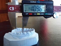
Methods: The study casts were retrieved from the orthodontic units of University College Hospital, Ibadan, and Military Hospital, Lagos. Sociodemographic variables, the mesiodistal width of the LM1, number of buccal grooves, and location of the buccal groove along the mesiodistal width of the LM1 were ascertained. Data were analysed using the SPSS software version 22. Paired t-test was used to assess the relationships between quantitative variables while the Chi-square test assessed qualitative variables and the level of significance was set at P < 0.05.
Results: The mean age of the patients was 15.50 ± 7.09 years. The mean mesiodistal widths of the lower right and left molars were 11.27 ± 0.78 mm and 11.41 ± 0.86 mm, respectively. Paired t-test showed that the left buccal groves were more anteriorly located than the right buccal grooves (P < 0.001). The buccal grooves were more anteriorly placed irrespective of the number of grooves present on the LM1, both left and right (P < 0.001).
Conclusion: The most appropriate nomenclature for the buccal groove of the LM1 is the anterior buccal groove. Caution must be exercised in classifying individuals with uncommon buccal groove location in clinical orthodontic practice.
Downloads
Article Details
The journal grants the right to make small numbers of printed copies for their personal non-commercial use under Creative Commons Attribution-Noncommercial-Share Alike 3.0 Unported License.
References
1. Adeyemi TA, Isiekwe MC. Comparing permanent tooth sizes of Nigerians and American Negroes. West Afr J Med 2003;22:63‑6.
2. Santoro M, Ayoub ME, Pardi VA, Cangialosi TJ. Mesiodistal crown dimensions and tooth size discrepancy of the permanent dentition of
Dominican Americans. Angle Orthod 2000;70:303‑7.
3. Scott RG. Dental morphology. In: Katzenberg MA, Saunders SR, editors. Biological Anthropology of the Human Skeleton. 2nd ed.
Hoboken, New Jersey: John Wiley and Sons, 2008; 265‑98.
4. Nelson SJ, Ash MM. The permanent mandibular molars. In: Dolan JJ, Loehr BS, editors. Wheeler’s Dental Anatomy, Physiology
and Occlusion. 9th ed. St. Louis, Missouri: Saunders/Elsevier, 2010; 189‑207.
5. Berkovitz BK, Holland GR, Moxham BJ. Dento‑osseous structures. In: Taylor A, Stader L, editors. Oral Anatomy, Histology and Embryology. 4th ed. Toronto: Mosby/Elsevier, 2009; 8‑61.
6. Goyal S, Goyal S. Pattern of dental malocclusion in orthodontic patients in Rwanda: A retrospective hospital based study. Rwanda Med
J 2012;69:13‑8.
7. Jang SY, Kim M, Chun YS. Differences in molar relationships and occlusal contact areas evaluated from the buccal and lingual aspects
using 3‑dimensional digital models. Korean J Orthod 2012;42:182‑9.
8. Hassan R, Rahimah AK. Occlusion, malocclusion and method of measurements – An overview. Arch Orofac Sci 2007;2:3‑9.
9. Guo Y, Han X, Xu H, Ai D, Zeng H, Bai D. Morphological characteristics influencing the orthodontic extraction strategies for
Angle’s class II division 1 malocclusions. Prog Orthod 2014;15:44.
10. Hashim HA, Al‑Ghamdi S. Tooth width and arch dimensions in normal and malocclusion samples: An odontometric study. J Contemp Dent Pract 2005;6:36‑51.
11. Sonika V, Harshaminder K, Madhushankari GS, Sri Kennath JA. Sexual dimorphism in the permanent maxillary first molar: A study
of the Haryana population (India). J Forensic Odontostomatol 2011;29:37‑43.
12. Eigbobo JO, Sote EO, Oredugba FA. Variations of crown dimensions of permanent dentitions in a selected population of Nigerian children. Nig Q J Hosp Med 2011;21:163‑8.
13. Rastogi P, Jain A, Kotian S, Rastogi S. Sexual dimorphism – An odontometric approach. Anthropology 2013;1:104.
14. Bunger E, Jindal R, Pathania D, Bunger R. Mesiodistal crown dimensions of the permanent dentition among school going children
in Punjab population: An aid in sex determination. Int J Dent Health Sci 2014;1:13‑23.
15. Alatrach AB, Saleh FK, Osman E. The prevalence of malocclusion and orthodontic treatment need in a sample of Syrian children. Eur
Sci J 2014;10:230‑47.


